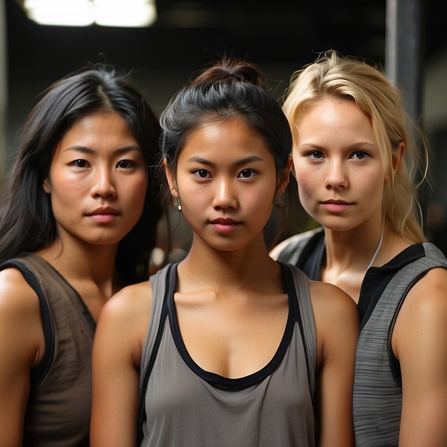
Ears
8 structured phenotype dimensions · drawn from peer-reviewed scales
Ears
General Description: Ears are sensory organs involved in hearing and maintaining balance. They come in various shapes and sizes and are often unique to each individual.
Ethnic Variations: Ear size, shape, and lobe attachment can vary across different ethnicities.
Cultural Significance: Ears have been adorned with various forms of jewelry in many cultures, reflecting aesthetic and societal norms.
AI Character Design Considerations: Including a range of ear shapes and sizes in AI character design adds to the diversity and detail of the models.
Ears — taxonomy
8 dimensions · 8 photo-assessable · v1.0.0 · UBERON: UBERON:0001690
External ear anatomy: helix, antihelix, tragus, lobule, ear axis, projection. Dimensions are drawn from craniofacial anthropometry (Farkas) and otoplasty literature (McDowell, Mustardé, Furnas). The earlobe attachment dimension (free/attached/intermediate) is the canonical Mendelian-genetics teaching example, though contemporary research has shown the trait is more polygenic and continuous than the textbook treatment suggests.
Dimensions
Helix morphology
photo-observablecategorical · helix_qualitative
Curvature and prominence of the helix (the outer rim of the ear).
Aligned with otoplasty-literature descriptors of helical-rim morphology.
Valid values (5)
well_curvedWell-curved— Smooth, continuous helical curve from superior to lobule.flattened_superiorFlattened superiorly— Reduced curve at the superior helix; common otoplasty correction target.stahls_earStahl's ear— Third crus visible; abnormal cartilage fold creating a pointed superior helix.cup_ear_constrictedCup ear / constricted— Furled or rolled helical rim creating a cupped appearance.asymmetricAsymmetric— Notable left-right helical morphology difference.
Antihelix definition
photo-observableordinal · antihelix_qualitative
Definition of the antihelical fold. Loss of antihelix definition is the primary cause of prominent ears.
Mustardé JC (1963). The correction of prominent ears using simple mattress sutures. British Journal of Plastic Surgery, 16. Antihelix-fold definition is the central otoplasty-correction landmark.
Valid values (3)
well_definedWell-defined— Clear antihelical fold; ear lies close to head.softly_definedSoftly defined— Antihelix visible but fold not crisp.absent_unfurledAbsent / unfurled— Antihelical fold absent or substantially reduced; ear appears to project from head.
Ear protrusion (auriculo-cephalic angle)
photo-observableordinal · auriculocephalic_angle_categorical
Angle between the ear and the side of the head, captured ordinally.
Furnas DW (1968). Correction of prominent ears by concha-mastoid sutures. Plastic and Reconstructive Surgery, 42(3). Reference: auriculo-cephalic angle ~20-25° aesthetic norm.
Valid values (4)
flat_against_headFlat against head— Auriculo-cephalic angle below ~15°; ear barely projects from head.normalNormal— Angle approximately 15-25°.prominentProminent— Angle 25-35°; visibly prominent.very_prominentVery prominent— Angle >35°; pronounced ear protrusion ('lop ear' / 'cup ear' in extreme cases).
Earlobe attachment
photo-observablecategorical · earlobe_attachment_continuum
Degree of attachment of the earlobe to the side of the head. Classically taught as two-state (free/attached) but contemporary genetics work demonstrates a continuous distribution; three buckets capture the meaningful variation.
Shaffer JR et al. (2017). Genome-wide association study reveals multiple loci influencing normal human earlobe attachment. American Journal of Human Genetics, 101(6). The classic Mendelian-genetics two-state classification is now understood as a polygenic continuum; this scale uses three buckets reflecting that continuum.
Valid values (3)
freeFree / detached— Earlobe hangs free of the head; clear gap between lobule and adjacent skin.intermediateIntermediate— Partial attachment; lobule attaches to head along part of its edge but not fully.attachedAttached— Lobule attaches to head along its entire posterior edge; no free hanging.
Earlobe size
photo-observableordinal · lobule_size_qualitative
Relative size of the earlobe.
Aligned with descriptors used in otoplasty and forensic-anthropology literature.
Valid values (4)
smallSmall— Limited lobule mass; minimal hanging tissue.mediumMedium— Average lobule size.largeLarge— Pronounced lobule mass; large hanging tissue.stretchedStretched (gauge / piercing)— Lobule visibly stretched from gauge piercings or repeated heavy earrings; native size not assessable.
Tragus morphology
partly photo-observablecategorical · tragus_qualitative
Shape of the tragus (the small projection anterior to the ear canal).
Otologic anatomy descriptors aligned with otologic-surgery references.
Valid values (4)
single_pointedSingle-pointed— One distinct tragal projection.double_pointedDouble-pointed— Two visible projections; common variant.roundedRounded— Tragus visible but rounded rather than pointed.minimalMinimal— Tragus barely visible; small or recessed.
Ear axis (vertical inclination)
partly photo-observablecategorical · ear_inclination_categorical
Inclination of the long ear axis relative to vertical, viewed laterally.
Farkas LG (1994). Anthropometry of the Head and Face, 2nd Edition. Reference: ear axis approximately 15-20° posterior tilt from vertical.
Valid values (3)
verticalVertical / minimal tilt— Ear axis nearly vertical.moderate_posterior_tiltModerate posterior tilt (~15-25°)— Within Farkas reference range.marked_posterior_tiltMarked posterior tilt (>25°)— Pronounced backward inclination.
Overall ear size
photo-observableordinal · ear_length_qualitative
Overall ear length proportional to face.
Farkas LG (1994). Reference: total ear height approximately equal to nasal length in European-population aesthetic norm.
Valid values (3)
smallSmall— Ear length below population median.mediumMedium— Average ear length.largeLarge— Ear length above population median; common with aging (ears continue growing through life).
References (5)
- Farkas LG (1994). Anthropometry of the Head and Face, 2nd Edition. Raven Press.
- Mustardé JC (1963). The correction of prominent ears using simple mattress sutures. British Journal of Plastic Surgery, 16: 170-178.
- Furnas DW (1968). Correction of prominent ears by concha-mastoid sutures. Plastic and Reconstructive Surgery, 42(3): 189-193.
- Shaffer JR, Li J, Lee MK, et al. (2017). Genome-wide association study reveals multiple loci influencing normal human earlobe attachment. American Journal of Human Genetics, 101(6): 913-924.
- McDowell AJ (1968). Goals in otoplasty for protruding ears. Plastic and Reconstructive Surgery, 41(1).
Top-coverage ethnic groups
Groups with the most image-grounded phenotype data — sorted by Data Depth score
- Soninken=39 · 85/100
- Tatarsn=70 · 85/100
- Uzbeksn=59 · 85/100
- Tuluvasn=52 · 84/100
- Irishn=49 · 84/100
- Iranunn=48 · 83/100
- Makassaresen=46 · 83/100
- Icelandersn=57 · 83/100
- Igbon=52 · 82/100
- Welshn=66 · 82/100
- Ibann=39 · 80/100
- Belarusiansn=62 · 80/100
- Ga-Adangben=35 · 79/100
- Estoniansn=73 · 79/100
- Javanesen=72 · 79/100
- Minangkabaun=51 · 79/100
- Mandinkan=54 · 79/100
- Tajiksn=37 · 79/100
- Ossetiansn=33 · 78/100
- Kadazan-Dusunn=33 · 78/100
- Kikuyun=34 · 78/100
- Garhwalisn=41 · 78/100
- Susun=26 · 77/100
- Tigrayansn=60 · 76/100